Seeing is believing!
Microscopy is more than just another method in science. The visualization of cellular and molecular structures offers scientists an insight into life on its most basic level and, thereby, a direct understanding in a very aesthetic way.
Our group is especially dedicated to apply high-end microscopy to deepen the understanding of infectious and immunological processes in human lung tissue and isolated pulmonary cells. We are using techniques such as spectral confocal or superresolution microscopy starting from static morphological 2D or 3D reconstruction up to functional and multi-dimensional live-cell or live-tissue imaging. This includes e.g. spectral FRET of mitochondrial ATP, Ca2+ or intracellular pH changes, induction of cell death pathways or organelle trafficking etc.
With the following images, we are presenting some examples of different microscopic techniques and structures of cells and tissue to illustrate our work.
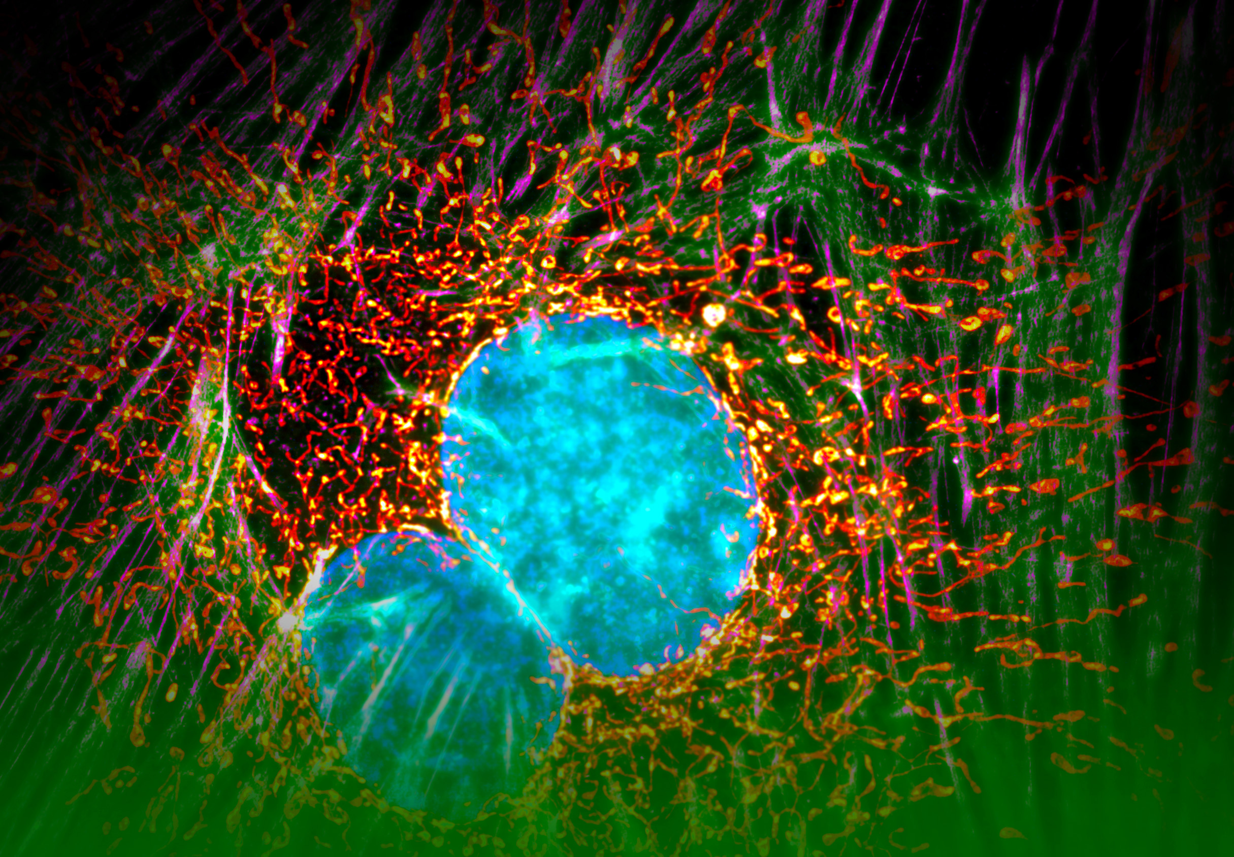
This image is a combination of conventional wide field imaging and the superresolution technique „structured illumination“ (max. res. 110nm). The hazy green colors are wide field actin fibers whereas the magenta stains the same by superresolution. Similarly, the nuclear DNA is stained in blue and cyan whereas the mitochondrial network is shown in red and yellow.
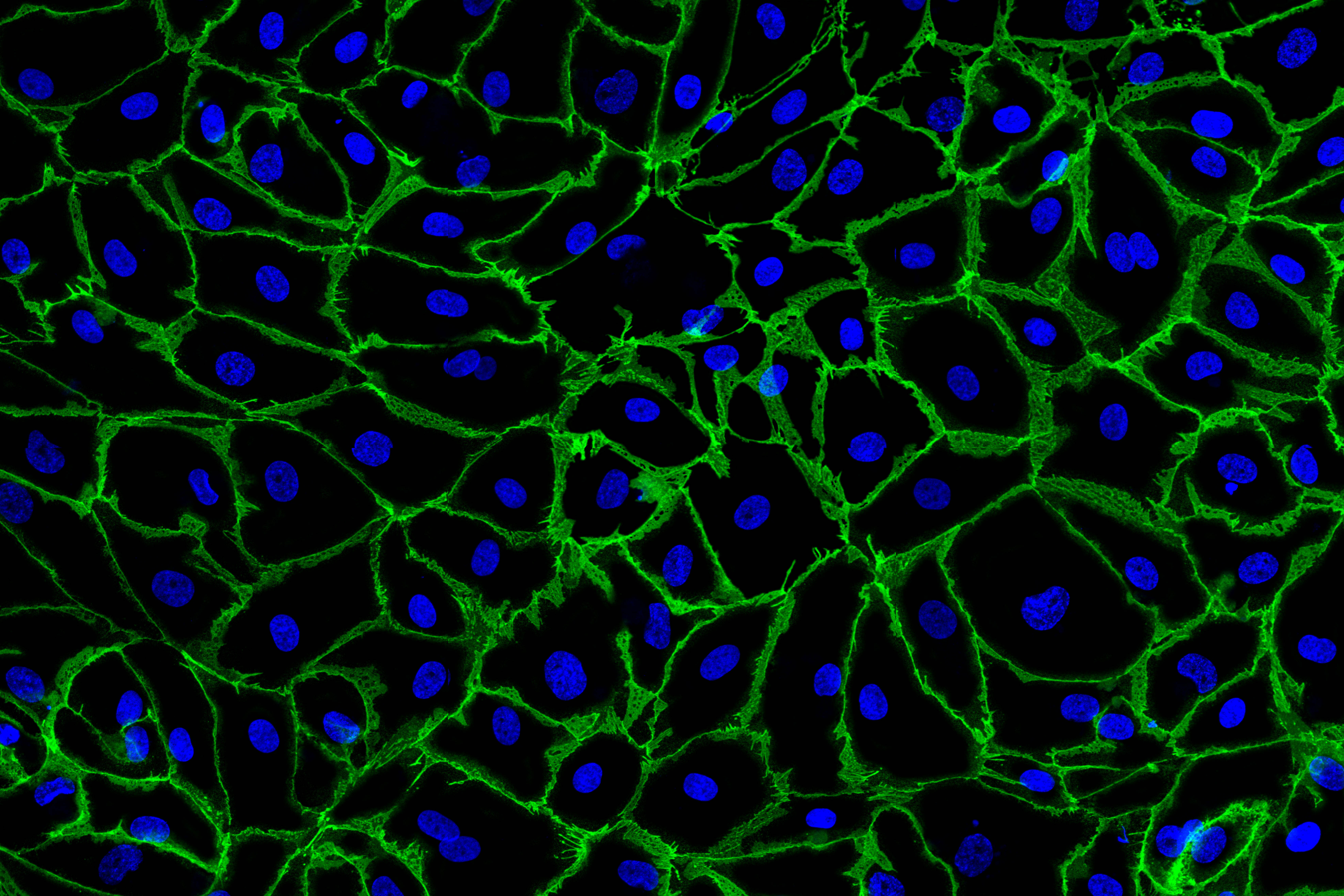
Already a conventional two-color confocal microscopy can demonstrate how cells organize in confluent monolayers. Here, endothelial cells are shown to express the junctional protein VE-Cadherin in green, which contributes to the sealing of vessels and capillaries to prevent edema formation.
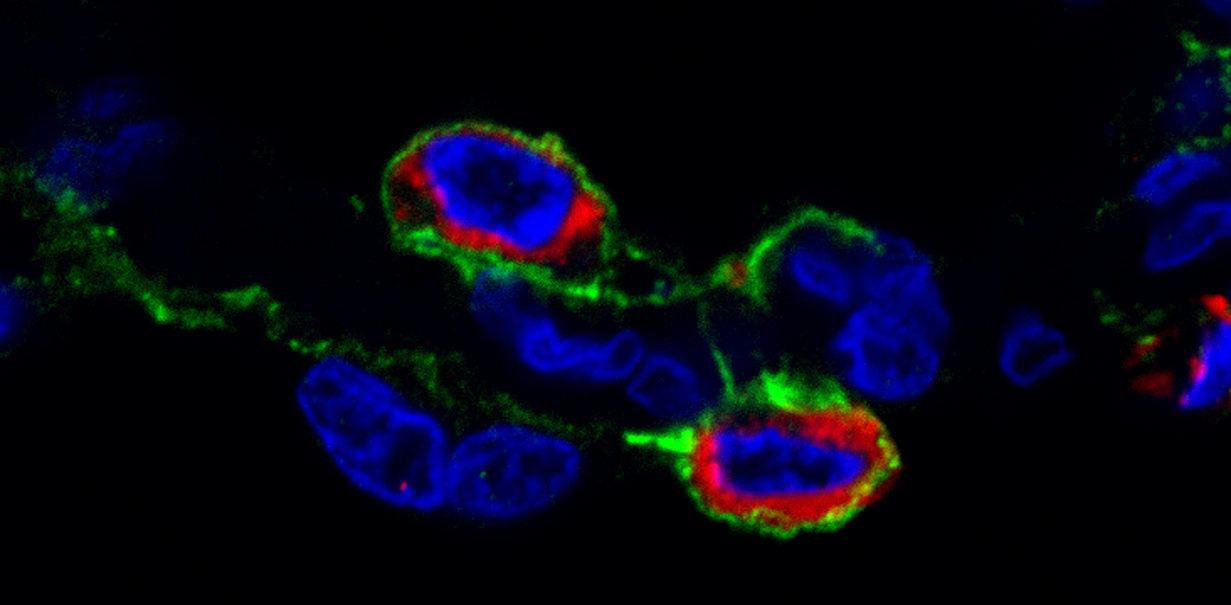
Next to the junctional proteins shown above, the alveolar homeostasis and fluid flux is regulated by additional enzymes such as the NaK-ATPase. This image is a spectral confocal microscopy of this protein (green) in the alveolar compartment of human lungs. It is localized on the basal membrane between alveolar epithelial type I cells and the capillary endothelium as well as surrounds the alveolar type II cells shown here by pro-surfactant protein C staining in red. Blue are again the nuclei.
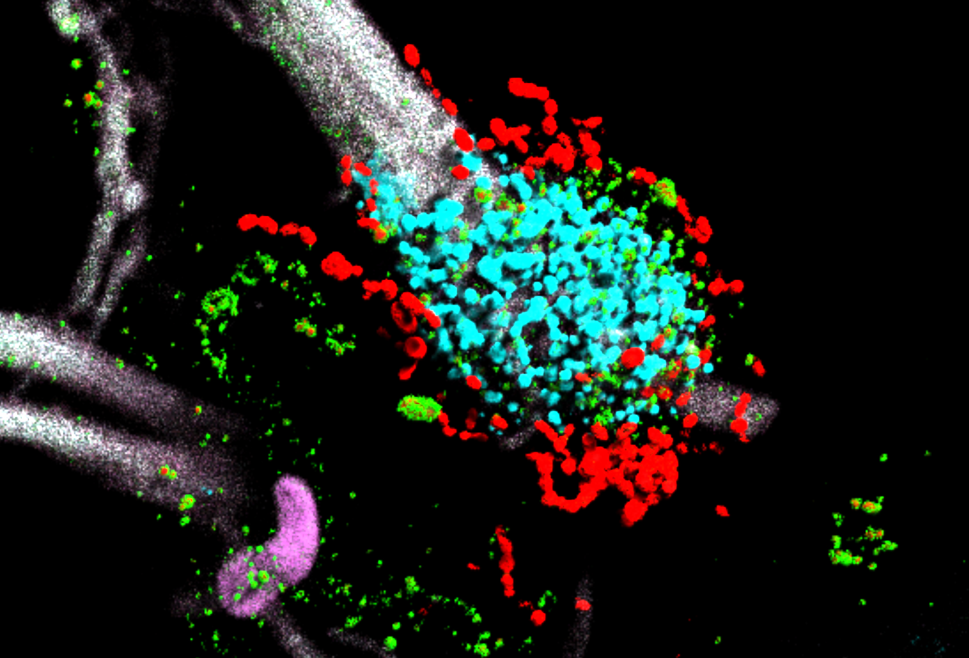
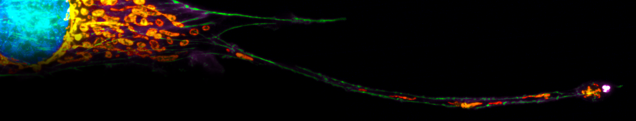
The alveolar epithelial cells, especially the type II cells, can be severely damaged by a bacterial attack occurring during pneumonia. This is shown here in a combinatory approach of spectral life-tissue imaging of the human alveolar compartment and an additional antibody staining after fixation. Streptococcus pneumoniae (the major pathogen causing pneumonia) is shown in red, while its important virulance factor pneumolysin (green) is liberated and inserts into the membranes of a mitochondrial (cyan) rich type II cell leading to cell death (nuclear magenta of caspase 3/7 activation as example here in a neighboring cell). The strong auto-fluorescence emission of the collagen backbone was unmixed by a linear fitting algorithm and is shown in white.